How to prepare for an ECG holter?

There are numerous diagnostic methods used by cardiologists in the process of detecting heart problems, and Holter ECG is one of the most commonly used.

Using this method, it is possible to obtain valuable information about the way the patient’s heart is functioning, whether it skips or accelerates while he is engaged in normal daily activities.

If you or someone close to you needs to visit a cardiologist soon for this reason, you will surely be interested in how to prepare for a Holter ECG.

What is an ECG of the heart?

An EKG or electrocardiogram is a painless, safe and non-invasive diagnostic method in which the heart’s electrical signals are recorded on paper or viewed on a computer.

An EKG is usually done in the doctor’s office and the recording does not take long, so it is sometimes difficult to notice deviations in the work of the heart, because it may happen that the problem is not active at that moment.

What is a Holter ECG?

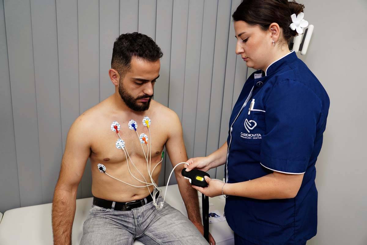
A Holter is actually a device the size of a mobile phone, which is connected to electrodes by wires and its task is to record the electrical activity of the heart for a long period of time, from 24 hours to 72 depending on the need.

Holter ECG of the heart is a painless, safe, non-invasive diagnostic method, so patients should not be afraid if it is recommended to them.

The doctor will suggest this method to the patient if the ordinary EKG cannot detect irregularities in the work of the heart and a longer time is needed to record and monitor the activity of the heart.

The most common candidates for a holter are patients who at certain times feel that their heart is working irregularly or have fainting spells or some other symptoms that raise the suspicion of the existence of heart disease.

When does a doctor recommend a heart holter?

There are numerous reasons why your doctor may recommend this diagnostic method. Among the most common are:

  • palpitations
  • heart palpitations
  • rapid pulse
  • dizziness
  • fainting
  • loss of consciousness
  • loss of breath
  • chest pain

All these symptoms actually belong to the symptoms of cardiovascular diseases and therefore it is very important that doctors first check the condition of the heart.

Patients who have had a heart attack or have some kind of heart disease are often recommended a holter monitor so that the cardiologist can have insight into the condition of the heart and how it is responding to therapy.

Monitoring the effects of therapy in people with diagnosed arrhythmias also cannot be effective without a cardiac holter.

Candidates for this method in certain time intervals will also be people with an implanted pacemaker, because it is necessary to check the operation and efficiency of the pacemaker.

How to prepare for Holter?

By reading the previous lines, you have already done a lot of preparation. You saw the advantages of this diagnostic method and learned that it is non-invasive, safe and painless!

This is very important to access this method easily.

Before putting the device on, you should take a bath, as you will not be able to do this while you have the heart holter on your body. It’s not hard to take off, but you’ll hardly be able to put it back on properly after a shower.

A trained technician will insert the Holter and remove the device when you return to the office after 24 hours or more, depending on the cardiologist’s instructions.

Men often have to shave their chest area so the electrodes can be attached to the body.

After the electrodes are placed, they are connected to the device by wires, which you can fasten around your waist. Holter and electrodes will not even be noticed under clothes.

If necessary, the technician should explain to you any doubts you have regarding the use of the device, because it is in everyone’s interest that the recording is successful.

While wearing the halter it is necessary to keep an activity log. You should write down what you did during which period, and especially you should write down and describe if you feel any discomfort. By comparing the diary and the information from the holter, the cardiologist will get a bigger picture of your condition.

What to expect after Holter?

After your Holter ECG is removed, your doctor analyzes the results and your activity log and makes a diagnosis or refers you for further tests.

Sometimes the data obtained in this way will be sufficient for a diagnosis, so you will be recommended therapy and the frequency of cardiac controls.
Ultrasound of the heart: What does it reveal and how often should it be done?

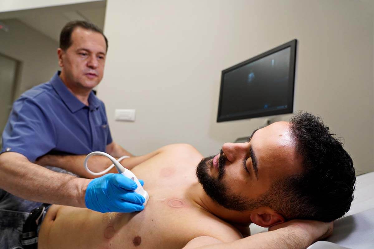
Echocardiography is an ultrasound test that checks the structure and function of the heart. In this way, you can see the shape, size and function of the heart, its chambers and atria, the location and dimensions of the damage, whether there is fluid around the heart, how the heart pumps blood and the appearance of the valves. A heart ultrasound usually takes up to 30 minutes. Ultrasound rays are not harmful, so children and pregnant women can be exposed to them.

What is an ultrasound of the heart (ECHO of the heart)?

Echocardiography is a graphic representation of the movement and work of the heart. A doctor often combines an echo with a Doppler ultrasound to assess blood flow through the heart valves. Echocardiography does not use radiation, which is an advantage over other diagnostic methods such as x-rays and CT scans, which involve the use of small amounts of radiation.

What can be detected by echocardiography?

Echocardiography can detect many types of heart disease, such as:

  • Cardiomyopathy. A disease of the heart muscle that is manifested by changes in the heart muscle such as thinning, thickening or reduced elasticity.
  • Cardiac arrhythmia. An ultrasound scan may be used to assess whether the heart has a regular heartbeat.
  • Infective endocarditis. Endovascular infection of cardiovascular structures (usually heart valves, but sometimes large blood vessels).
  • Heart failure (heart failure). A condition in which the heart muscle is weakened and cannot pump blood efficiently to the organs. This can cause fluid build-up (congestion) in the blood vessels and lungs and edema (swelling) in the feet and other parts of the body.
  • Pericarditis – Inflammation or infection of the pericardium.
  • Pericardial effusion or tamponade. A condition caused by the accumulation of fluid in the pericardial space. This puts pressure on the heart muscle, which cannot beat and pump blood normally. The condition is life-threatening.
  • Defects of the walls of the atrium or septum. These defects occur in the upper atria or in the lower ventricles (ventricles). The condition leads to heart failure or poor blood flow. They belong to the most common congenital heart defects.
  • Diseases of heart valves. Malfunction of one or more heart valves that can cause disruption of blood flow to the heart. An echocardiogram can also check for an infection of the heart valve tissue.
  • Aneurysm. Enlargement and weakening of part of the heart muscle or the aorta (the large artery that carries oxygenated blood from the heart to the rest of the body). An aneurysm may be at risk of rupture.
  • Congenital heart diseases. Defects in one or more structures of the heart that occur during fetal development, such as a ventricular septal defect (a hole in the wall between the two lower chambers of the heart).

How often should a heart ultrasound be done?

Heart screenings, which include ultrasound, should begin around age 25. It is recommended that examinations, if there are no complaints or risk of hereditary diseases, be done every two to four years.

How often the echocardiogram should be repeated depends on the findings of the initial test. If the result of the initial echocardiogram was normal, there is no need to repeat it except in case of new complaints. Heart failure and valvular heart disease are the two most common diseases that require more frequent heart ultrasound examinations.

Depending on the severity of the disease, the echocardiogram may need to be repeated more often. For example, a more severe form of aortic valve stenosis usually needs monitoring every 6-12 months; and for a milder form, the control is three years. The same rule applies to monitoring other valvular pathologies.

Why is it important to maintain normal blood pressure?

Blood pressure is the pressure of the blood in the heart’s arteries. Arteries carry blood from the heart to all organs. Blood pressure normally rises and falls throughout the day. Average blood pressure is not the same for men and women, and tends to increase with age in both sexes. According to the European Heart Association, the optimal (ideal) blood pressure for adults (over 20 years old) is less than 120/80 mmHg, and normal blood pressure is considered up to 139/89 mmHg.

Why is blood pressure measurement important?

Blood pressure measurement is quick and painless. That is why it is important for people with normal values to measure it occasionally at home. High blood pressure is called the “silent killer” because there are usually no warning signs or symptoms, and many people don’t know they have it.

Before age 55, men are more likely to develop high blood pressure. Women are more likely to develop high blood pressure after menopause.

For people who have elevated blood pressure values, it is important to have regular control and measurement of blood pressure by a cardiologist who will give instructions and possibly prescribe drug therapy.

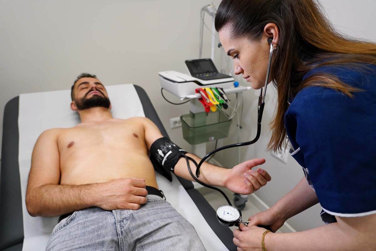
The pressure is usually measured on the upper arm of both hands. The patient should be seated and relaxed for at least five minutes before taking the blood pressure. Two to three values are measured with an interval of 2-3 minutes. The physiological difference between the two hands is up to 10 mmHg. The official pressure value is the one that was higher by measurement.

What is upper and lower blood pressure?

Blood pressure is recorded through two numbers:

Systolic blood pressure – shows how much pressure the blood exerts on the walls of the arteries when the heart beats.

Diastolic blood pressure – shows how much pressure the blood exerts on the walls of the arteries while the heart is resting between beats.

Usually more attention is paid to systolic blood pressure (the first number) as the main risk factor for cardiovascular disease in people over 50 years of age. In most people, systolic blood pressure increases with age due to increased stiffness of the large arteries. However, the diagnosis of high blood pressure can be established if there is an increase in the value of both the upper and lower blood pressure. According to recent research, the risk of death from coronary heart disease and stroke doubles for every 20 mmHg increase in systolic or 10 mmHg increase in diastolic blood pressure in people aged 40 to 89 years.

How to maintain normal pressure values?

Many people with high blood pressure can lower their blood pressure or keep it within normal limits by making lifestyle changes.

This implies the following measures:

  • At least 150 minutes of physical activity per week (about 30 minutes a day, 5 days a week)
  • Leaving cigarettes if the person is a smoker
  • Healthy diet, limiting sodium (salt) and alcohol
  • Maintaining a healthy weight and diet in overweight people
  • Stress management