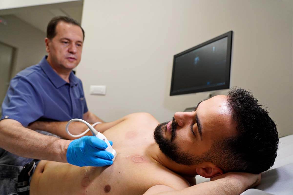
Echocardiography is an ultrasound test that checks the structure and function of the heart. In this way, you can see the shape, size and function of the heart, its chambers and atria, the location and dimensions of the damage, whether there is fluid around the heart, how the heart pumps blood and the appearance of the valves. A heart ultrasound usually takes up to 30 minutes. Ultrasound rays are not harmful, so children and pregnant women can be exposed to them.
What is an ultrasound of the heart (ECHO of the heart)?
Echocardiography is a graphic representation of the movement and work of the heart. A doctor often combines an echo with a Doppler ultrasound to assess blood flow through the heart valves. Echocardiography does not use radiation, which is an advantage over other diagnostic methods such as x-rays and CT scans, which involve the use of small amounts of radiation.
What can be detected by echocardiography?
Echocardiography can detect many types of heart disease, such as:
- Cardiomyopathy. A disease of the heart muscle that is manifested by changes in the heart muscle such as thinning, thickening or reduced elasticity.
- Cardiac arrhythmia. An ultrasound scan may be used to assess whether the heart has a regular heartbeat.
- Infective endocarditis. Endovascular infection of cardiovascular structures (usually heart valves, but sometimes large blood vessels).
- Heart failure (heart failure). A condition in which the heart muscle is weakened and cannot pump blood efficiently to the organs. This can cause fluid build-up (congestion) in the blood vessels and lungs and edema (swelling) in the feet and other parts of the body.
- Pericarditis – Inflammation or infection of the pericardium.
- Pericardial effusion or tamponade. A condition caused by the accumulation of fluid in the pericardial space. This puts pressure on the heart muscle, which cannot beat and pump blood normally. The condition is life-threatening.
- Defects of the walls of the atrium or septum. These defects occur in the upper atria or in the lower ventricles (ventricles). The condition leads to heart failure or poor blood flow. They belong to the most common congenital heart defects.
- Diseases of heart valves. Malfunction of one or more heart valves that can cause disruption of blood flow to the heart. An echocardiogram can also check for an infection of the heart valve tissue.
- Aneurysm. Enlargement and weakening of part of the heart muscle or the aorta (the large artery that carries oxygenated blood from the heart to the rest of the body). An aneurysm may be at risk of rupture.
- Congenital heart diseases. Defects in one or more structures of the heart that occur during fetal development, such as a ventricular septal defect (a hole in the wall between the two lower chambers of the heart).
How often should a heart ultrasound be done?
Heart screenings, which include ultrasound, should begin around age 25. It is recommended that examinations, if there are no complaints or risk of hereditary diseases, be done every two to four years.
How often the echocardiogram should be repeated depends on the findings of the initial test. If the result of the initial echocardiogram was normal, there is no need to repeat it except in case of new complaints. Heart failure and valvular heart disease are the two most common diseases that require more frequent heart ultrasound examinations.
Depending on the severity of the disease, the echocardiogram may need to be repeated more often. For example, a more severe form of aortic valve stenosis usually needs monitoring every 6-12 months; and for a milder form, the control is three years. The same rule applies to monitoring other valvular pathologies.


